OpenPlant Coverage

The new OpenPlant toolkit for Marchantia, a platform for basic research and plant synthetic biology applications

The new OpenPlant toolkit for Marchantia, a platform for basic research and plant synthetic biology applications

Systematic tools for reprogramming plant gene expression in a simple model, Marchantia polymorpha.

Sauret-Güeto S, Frangedakis E, Silvestri L, Rebmann M, Tomaselli M, Markel K, Delmans M, West A, Patron NJ, Haseloff J.

ACS Synth. Biol. 2020, 9, 4, 864–882

https://doi.org/10.1021/acssynbio.9b00511

Combining Transient Expression and Cryo-EM to Obtain High-Resolution Structures of Luteovirid Particles

OpenPlant PI George Lomonossoff and colleagues published their work on combining transient expression and cryo-EM to obtain high-resolution structures of luteovirid particles:

Byrne et al. (2019) Graphical abstract

Byrne et al. (2019) Graphical abstract

Combining Transient Expression and Cryo-EM to Obtain High-Resolution Structures of Luteovirid Particles

Matthew J. Byrne, John F.C.Steele, Emma L. Hesketh, Miriam Walden, Rebecca F. Thompson, George P. Lomonossoff, and Neil A. Ranson

Structure (2019) https://doi.org/10.1016/j.str.2019.09.010

Abstract:

The Luteoviridae are pathogenic plant viruses responsible for significant crop losses worldwide. They infect a wide range of food crops, including cereals, legumes, cucurbits, sugar beet, sugarcane, and potato and, as such, are a major threat to global food security. Viral replication is strictly limited to the plant vasculature, and this phloem limitation, coupled with the need for aphid transmission of virus particles, has made it difficult to generate virus in the quantities needed for high-resolution structural studies. Here, we exploit recent advances in heterologous expression in plants to produce sufficient quantities of virus-like particles for structural studies. We have determined their structures to high resolution by cryoelectron microscopy, providing the molecular-level insight required to rationally interrogate luteovirid capsid formation and aphid transmission, thereby providing a platform for the development of preventive agrochemicals for this important family of plant viruses.

Improving plant transient expression through the rational design of synthetic 5′ and 3′ untranslated regions

Dr. Heydrien Peyret, Prof. James Brown and OpenPlant PI Prof. George Lomonossoff published their work on improving plant transient expression through the rational design of synthetic 5' and 3' untranslated regions in expression vectors:

Peyret et al. (2019): “Fig 1: The Synth expression cassette.”

Peyret et al. (2019): “Fig 1: The Synth expression cassette.”

Improving plant transient expression through the rational design of synthetic 5′ and 3′ untranslated regions

Hadrien Peyret, James K. M. Brown & George P. Lomonossoff

Plant Methods Volume 15, Article number: 108 (2019)

https://plantmethods.biomedcentral.com/articles/10.1186/s13007-019-0494-9

Abstract:

Background

The growing field of plant molecular farming relies on expression vectors that allow high yields of recombinant proteins to be produced through transient gene expression. While numerous expression vectors currently exist for this purpose, there are very few examples of systematic efforts to improve upon these. Moreover, the current generation of expression systems makes use of naturally-occurring regulatory elements, typically selected from plant viruses, to maximise yields. This study aims to use rational design to generate synthetic sequences that can rival existing ones.

Results

In this work, we present the rational design of novel synthetic 5′ and 3′ untranslated regions (UTRs) which can be used in various combinations to modulate accumulation levels of transiently-expressed recombinant proteins. Using the pEAQ-HT expression vector as a point of comparison, we show that pre-existing expression systems can be improved by the deployment of rationally designed synthetic UTRs. Notably, we show that a suite of short, synthetic 5′UTRs behave as expression enhancers that outperform the HT 5′UTR present in the CPMV-HT expression system. Furthermore, we confirm the critical role played by the 3′UTR of cowpea mosaic virus RNA-2 in the performance of the CPMV-HT system. Finally, we use the knowledge obtained from these results to develop novel expression vectors (named pHRE and pHREAC) that equal or outperform pEAQ-HT in terms of recombinant protein yield. These new vectors are also domesticated for the use of certain Type IIS restriction enzymes, which allows for quicker cloning and straightforward assessment of different combinations of UTRs.

Conclusions

We have shown that it is possible to rationally design a suite of expression modulators in the form of synthetic UTRs. We have created novel expression vectors that allow very high levels of recombinant protein expression in a transient expression context. This will have important consequences for future efforts to develop ever-better plant transient overexpression vectors for research or industrial applications.

New publications from the Baulcombe lab

OpenPlant PI David Baulcombe and colleagues recently published two papers: (1) on the miRNA-Argonaute machinery in the unicellular green alga Chlamydomonas reinhardtii, and (2) on the application of miRNAs for regulation of synthetic gene systems in this organism:

Chung et al. (2019): “Figure 1: Structural features of Chlamydomonas Argonautes.”

Chung et al. (2019): “Figure 1: Structural features of Chlamydomonas Argonautes.”

Distinct roles of Argonaute in the green alga Chlamydomonas reveal evolutionary conserved mode of miRNA-mediated gene expression

Betty Y.-W. Chung, Adrian Valli, Michael J. Deery, Francisco J. Navarro, Katherine Brown, Silvia Hnatova, Julie Howard, Attila Molnar & David C. Baulcombe

Sci Rep. 2019; 9: 11091. doi: 10.1038/s41598-019-47415-x

https://www.nature.com/articles/s41598-019-47415-x.pdf

Abstract:

The unicellular green alga Chlamydomonas reinhardtii is evolutionarily divergent from higher plants, but has a fully functional silencing machinery including microRNA (miRNA)-mediated translation repression and mRNA turnover. However, distinct from the metazoan machinery, repression of gene expression is primarily associated with target sites within coding sequences instead of 3′UTRs. This feature indicates that the miRNA-Argonaute (AGO) machinery is ancient and the primary function is for post transcriptional gene repression and intermediate between the mechanisms in the rest of the plant and animal kingdoms. Here, we characterize AGO2 and 3 in Chlamydomonas, and show that cytoplasmically enriched Cr-AGO3 is responsible for endogenous miRNA-mediated gene repression. Under steady state, mid-log phase conditions, Cr-AGO3 binds predominantly miR-C89, which we previously identifed as the predominant miRNA with efects on both translation repression and mRNA turnover. In contrast, the paralogue Cr-AGO2 is nuclear enriched and exclusively binds to 21-nt siRNAs. Further analysis of the highly similar Cr-AGO2 and Cr-AGO 3 sequences (90% amino acid identity) revealed a glycine-arginine rich N-terminal extension of ~100 amino acids that, given previous work on unicellular protists, may associate AGO with the translation machinery. Phylogenetic analysis revealed that this glycine-arginine rich N-terminal extension is present outside the animal kingdom and is highly conserved, consistent with our previous proposal that miRNA-mediated CDS-targeting operates in this green alga.

Navarro and Baulcombe (2019): “Figure 1: Construction of a synthetic circuit to measure miRNA-dependent gene repression.”

Navarro and Baulcombe (2019): “Figure 1: Construction of a synthetic circuit to measure miRNA-dependent gene repression.”

miRNA-mediated regulation of synthetic gene circuits in the green alga Chlamydomonas reinhardtii

Francisco J. Navarro and David C. Baulcombe

ACS Synth Biol. 2019 February 15; 8(2): 358–370. doi:10.1021/acssynbio.8b00393.

https://www.ncbi.nlm.nih.gov/pmc/articles/PMC6396871/pdf/emss-81902.pdf

Abstract:

microRNAs (miRNAs), small RNA molecules of 20–24 nts, have many features that make them useful tools for gene expression regulation — small size, flexible design, target predictability and action at a late stage of the gene expression pipeline. In addition, their role in fine-tuning gene expression can be harnessed to increase robustness of synthetic gene networks. In this work we apply a synthetic biology approach to characterize miRNA-mediated gene expression regulation in the unicellular green alga Chlamydomonas reinhardtii. This characterization is then used to build tools based on miRNAs, such as synthetic miRNAs, miRNA-responsive 3’UTRs, miRNA decoys and self-regulatory loops. These tools will facilitate the engineering of gene expression for new applications and improved traits in this alga.

Genome-wide transcription factor binding in leaves from C3 and C4 grasses

OpenPlant PI Julian Hibberd and colleagues published their work on the transcription factor binding repertoire associated with C3 and C4 photosynthesis:

Burgess et al. (2019): “Fig 6: Hyper-conserved cis-elements in grasses recruited into C4 photosynthesis.”

Burgess et al. (2019): “Fig 6: Hyper-conserved cis-elements in grasses recruited into C4 photosynthesis.”

Genome-wide transcription factor binding in leaves from C3 and C4 grasses

Steven J Burgess, Ivan Reyna-Llorens, Sean Ross Stevenson, Pallavi Singh, Katja Jaeger, and Julian M Hibberd

Plant Cell. 2019. pii: tpc.00078.2019. doi: 10.1105/tpc.19.00078.

http://www.plantcell.org/content/plantcell/early/2019/08/19/tpc.19.00078.full.pdf

Abstract:

The majority of plants use C3 photosynthesis, but over sixty independent lineages of angiosperms have evolved the C4 pathway. In most C4 species, photosynthesis gene expression is compartmented between mesophyll and bundle sheath cells. We performed DNaseI-SEQ to identify genome-wide profiles of transcription factor binding in leaves of the C4 grasses Zea mays, Sorghum bicolor and Setaria italica as well as C3 Brachypodium distachyon. In C4 species, while bundle sheath strands and whole leaves shared similarity in the broad regions of DNA accessible to transcription factors, the short sequences bound varied. Transcription factor binding was prevalent in gene bodies as well as promoters, and many of these sites could represent duons that impact gene regulation in addition to amino acid sequence. Although globally there was little correlation between any individual DNaseI footprint and cell-specific gene expression, within individual species transcription factor binding to the same motifs in multiple genes provided evidence for shared mechanisms governing C4 photosynthesis gene expression. Furthermore, interspecific comparisons identified a small number of highly conserved transcription factor binding sites associated with leaves from species that diverged around 60 million years ago. These data therefore provide insight into the architecture associated with C4 photosynthesis gene expression in particular and characteristics of transcription factor binding in cereal crops in general.

A Biomaker team has made it to the final of the BBSRC Innovator of the Year 2019 Awards

Close-up-cabinet-615x340.png

A Biomaker team with participants from Quadram Institute Bioscience (QIB), the Earlham Institute (EI), the John Innes Centre (JIC) and the University of Oxford, has developed a small scale speed breeding cabinet, which has qualified them for the final of the BBSRC Innovator of the Year 2019 Awards. Read more about this story here:

https://www.jic.ac.uk/news/norwich-research-park-team-in-line-for-early-career-innovator-award/

A specialized metabolic network selectively modulates Arabidopsis root microbiota

A specialized metabolic network selectively modulates Arabidopsis root microbiota

A specialized metabolic network selectively modulates Arabidopsis root microbiota.

Ancheng C. Huang, Ting Jiang, Yong-Xin Liu, Yue-Chen Bai, James Reed, Baoyuan Qu, Alain Goossens, Hans-Wilhelm Nützmann, Yang Bai, Anne Osbourn.

Science 10 May 2019: Vol. 364, Issue 6440

https://doi.org/10.1126/science.aau6389

Cas9-mediated mutagenesis of potato starch branching enzymes generates a range of tuber starch phenotypes

Cas9-mediated mutagenesis of potato starch branching enzymes generates a range of tuber starch phenotypes

Cas9-mediated mutagenesis of potato starch branching enzymes generates a range of tuber starch phenotypes.

Tuncel, A., Corbin, K.R., Ahn‐Jarvis, J., Harris, S., Hawkins, E., Smedley, M.A., Harwood, W., Warren, F.J., Patron, N.J. and Smith, A.M.

Plant Biotechnol J (2019) 17: 2259-2271.

https://doi.org/10.1111/pbi.1313

The protosteryl and dammarenyl cation dichotomy in polycyclic triterpene biosynthesis revisited: has this ‘rule’ finally been broken?

The protosteryl and dammarenyl cation dichotomy in polycyclic triterpene biosynthesis revisited: has this ‘rule’ finally been broken?

The protosteryl and dammarenyl cation dichotomy in polycyclic triterpene biosynthesis revisited: has this ‘rule’ finally been broken?

Michael J. Stephenson, Robert A. Field and Anne Osbourn.

Nat. Prod. Rep., 2019,36, 1044-1052

https://doi.org/10.1039/C8NP00096D

Two members of the DUF579 family are responsible for arabinogalactan methylation in Arabidopsis

Two members of the DUF579 family are responsible for arabinogalactan methylation in Arabidopsis

Two members of the DUF579 family are responsible for arabinogalactan methylation in Arabidopsis.

Henry Temple, Jenny C. Mortimer, Theodora Tryfona, Xiaolan Yu, Federico Lopez‐Hernandez, Mathias Sorieul, Nadine Anders, Paul Dupree

Plant Direct. 2019; 3: 1– 4.

https://doi.org/10.1002/pld3.117

OpenPlant Forum 2018: Engineering Plants for Bioproduction

Blog post by Dr Colette Matthewman

Over the past decade, synthetic biology has focussed much of its effort on microbial chassis as platform for bioproduction. The single cell simplicity and rapid life-cycles of these organisms, the prevalence of biological tools and the existing industry infrastructure for fermentation have made microbes a tempting playground for synthetic biologists wishing to make a range of chemicals and biomolecules, from flavours and fragrances to distributed manufacturing of highly complex metabolites for medicine, and an increasing number of companies are finding success in this arena (e.g. Ginkgo Bioworks, Amyris, Evolva, Antheia).

More recently, plants have been showing serious promise as viable production platforms for complex chemicals and biomolecules which in many cases simply can’t be made in single celled microbes. This year, the OpenPlant Forum explored some of the latest advances in plant bioproduction with inspiring talks from invited speakers and OpenPlant researchers highlighting a promising and exciting future for plant synthetic biology.

OpenPlant post-doc Ingo Appelhagen presents his work on anthocyanin pigment production in plant cell cultures.

OpenPlant post-doc Ingo Appelhagen presents his work on anthocyanin pigment production in plant cell cultures.

The first morning of the Forum focused on tools for refactoring regulation and simple test platforms for plant synthetic biology. Prof. Ian Small (University of Western Australia) opened the meeting with a keynote on the potential for using engineered RNA bonding proteins to control organelle gene expression. OpenPlant PI, Prof. Paul Dupree described research in his on engineering of polysaccharide structures in plants. We also had the first examples of plant production platforms: Dr Ingo Appelhagen presented his recently published work on the production of colourful anthocyanin molecules in plant cell cultures, while Dr Eva Thuenemann introduced the HyperTrans system developed in the Lomonossoff lab at the John Innes Centre for the transient expression of proteins in Nicotiana benthamiana, a wild relative of tobacco. Eva is working on plant-based production of a protein that could be used in a vaccine against East Coast Fever, a devastating disease in cattle in Africa. The HyperTrans platform is used by the Lomonossoff lab and recently established company Leaf Expression Systems to produce therapeutic proteins and virus-like particles for vaccines, including recent work on a new vaccine for the eradication of Polio.

The afternoon session explored the cutting edge in production of complex plant-derived natural products in yeast, with a keynote from Prof. Christina Smolke (Stanford University), followed with an insight into the engineering of triterpene production in N. benthamiana by Dr James Reed in the Osbourn lab (John Innes Centre), recently reviewed in Plant Cell Reports. These projects rely heavily on chemical and enzymatic biodiversity in nature. Dr Sam Brockington (University of Cambridge) talked about harnessing the global network of botanic gardens for access to plant diversity for metabolic engineering and synthetic biology, introducing a global database of living plant, seed and tissue collections called “Plant Search” – a perfect sedgeway into a panel discussion on Harnessing Global Biodiversity where Sam was joined by Dr Nicola Patron (Earlham Institute), Mr David Rejeski (Environmental Law Institute), and Dr Jenni Rant (SAW Trust). The discussions ranged from public opinion on synthetic biology (explored through the Global Garden workshop) and benefit sharing and dematerialisation, through to how blockchain (like the bitcoin) is being used in environmental contexts and whether blockchain technology trends can be applied to create/assign value for biodiversity.

Prof. Ralf Reski with his moss bioreactors

Prof. Ralf Reski with his moss bioreactors

Day two of the Forum continued on a theme of “Tools for Metabolic Engineering” with Prof. Claudia Vickers (University of Queensland) opening by introducing the Future Science Platform in Synthetic Biology that she leads at CSIRO, as well as numerous tools developed in her research lab. Claudia was followed by a trio of OpenPlant postdocs describing analysis to unravel the genetics of divergent metabolic pathways in Brassicaceae (Dr Zhenhua Liu), a search for new synthetic biology tools based on diversity of natural triterpene oxidation (Dr Michael Stephenson) and tools for engineering Marchantia’s chloroplasts (Dr Eftychis Frangedakis).

Moving on from the tools, we explored further plant-based bioproduction platforms, starting with an inspirational keynote from Prof. Ralf Reski (University of Freiburg) on the moss Physcomitrella patens that Ralf’s lab has established as a production platform for biopharmaceuticals, leading to foundation of the company Greenovation, which produces moss-aGal (agalsidase) for the treatment of Fabry disease, a rare but painful and potentially deadly disease. Subsequently, we heard from Prof. Alison Smith (University of Cambrige) about “Designer algae” and work towards predictable metabolic engineering in microalgae, and from Dr Eugenio Butelli (John Innes Centre) about the Tomato as a biofactory for making health promoting flavonoids.

The Forum was wrapped up for this year with a session on Sharing and Techno-Social Platforms, with an introduction from OpenPlant’s Prof Jim Haseloff, followed by Dr Linda Kahl (BioBricks Foundation) on the latest with the Open Material Transfer Agreement (Open MTA) which has been developed in collaboration with OpenPlant to enable sharing of DNA parts (publication coming soon!). Next up, Dr Joanne Kamens from not-for-profit plasmid distribution company, Addgene, revealed the freshly launched plant resource page and spoke about the upcoming adoption of the Open MTA as an option under which plasmids can be shared. Finally, Dr Richard Sever from bioRxiv spoke about preprint opportunities for synthetic biology.


Join us in Cambridge for the OpenPlant Forum 2019 | 29 – 31 July

Save the date!

Colour bio-factories: anthocyanin production in plant cell cultures

Colour bio-factories: Towards scale-up production of anthocyanins in plant cell cultures.

Appelhagen I, Wulff-Vester AK, Wendell M, Hvoslef-Eide AK, Russell J, Oertel A, Martens S, Mock HP, Martin C, Matros A (2018).

Metabolic Engineering. Volume 48, 2018, Pages 218-232

https://doi.org/10.1016/j.ymben.2018.06.004

DIY macrophotography and embracing the challenge of video documentation

Jennifer-Deegan.jpg

Dr Jennifer Deegan has been awarded an OpenPlant Fund grant to develop teaching materials to enable others to build duplicates of her focus stacking photography setup, and to capture images that can be used for teaching and publications in plant sciences. We caught up with her to find out what she has been up to and how her project is progressing.

Full details of her project can be found on the biomaker.org website.


Jennifer, please can you give a brief overview of your project?

Jennifer Deegan: The project follows on from my Biomaker 2017 project to build a low budget DIY Focus stacking photography system. The system takes photographs of tiny plant specimens about 2mm across, with the entire specimen in focus.

An image of a gametophyte fern, captured using the DIY Focus stacking photography system

An image of a gametophyte fern, captured using the DIY Focus stacking photography system

In the past it was not possible to take photographs of such tiny specimens and have them fully in focus. This was because single images taken at high magnification had only a very shallow depth of field. With this new technique we take about 40 photographs of a tiny specimen, with the camera moving progressively towards the subject. Then all of the focused parts of the images are cut out and amalgamated together into one fully focused image.

Commercial systems are available to do this, but they are very expensive. The more affordable ones only move the camera in increments of 2 micrometres. This is not small enough for use at very high magnification. Our system is very cheap and can moved in increments down to about 1/128th of a micrometre.

The DIY Focus stacking photography system

The DIY Focus stacking photography system

As part of this OpenPlant project we have two goals:

  • Document the construction of the focus stacking system so that others can copy it.
  • Use the system to take plant photos that have never before been possible. These photos will then be made available for plant science teaching and text books.


What inspired the project?

JD: I have always been frustrated that there are no great photos of fern gametophytes anywhere. Fern gametophytes have a very interesting planar heart shaped structure that is brought about by a tightly choreographed series of cell divisions. In the literature they are usually drawn by hand, because they are too small to be photographed in full focus. During my career break to raise my son, I have been working at home as a volunteer, to try to build a system that can take good, full focus, high magnification photographs of these structures.


What has been your favourite aspect of the project so far?

JD: The judges asked me to document my system using videos rather than just in writing. This threw me for a loop initially as I have never made video and didn't have the equipment. However, I have managed to cobble a system together, and am loving my new craft. The time, nuance and attention to detail that is needed to make a short video is amazing. The photo below shows the many photo, video and sound files that I had to record and line up in order to create one short video.  I'm now the proud owner of a YouTube channel. (You can visit it, and the other documentation on GitHub and Hackster via www.chlorophyllosophy.uk)

Editing videos that explain how the focus stacking system works

Editing videos that explain how the focus stacking system works


What are the biggest challenges you have come across?

JD: There have been a lot of challenges, particularly with the transition from written documentation to video.

The biggest problem is that my laptop is ten years old and is a bit slow for editing video. It cannot play my videos at full speed, so I have to upload them to YouTube between editing session to see what they look like. Saving the files out for upload to YouTube takes 2.5 hours for each video, so it is a slow process.

The DSLR filming the focus stacking setup, with decoy camera body in place

The DSLR filming the focus stacking setup, with decoy camera body in place

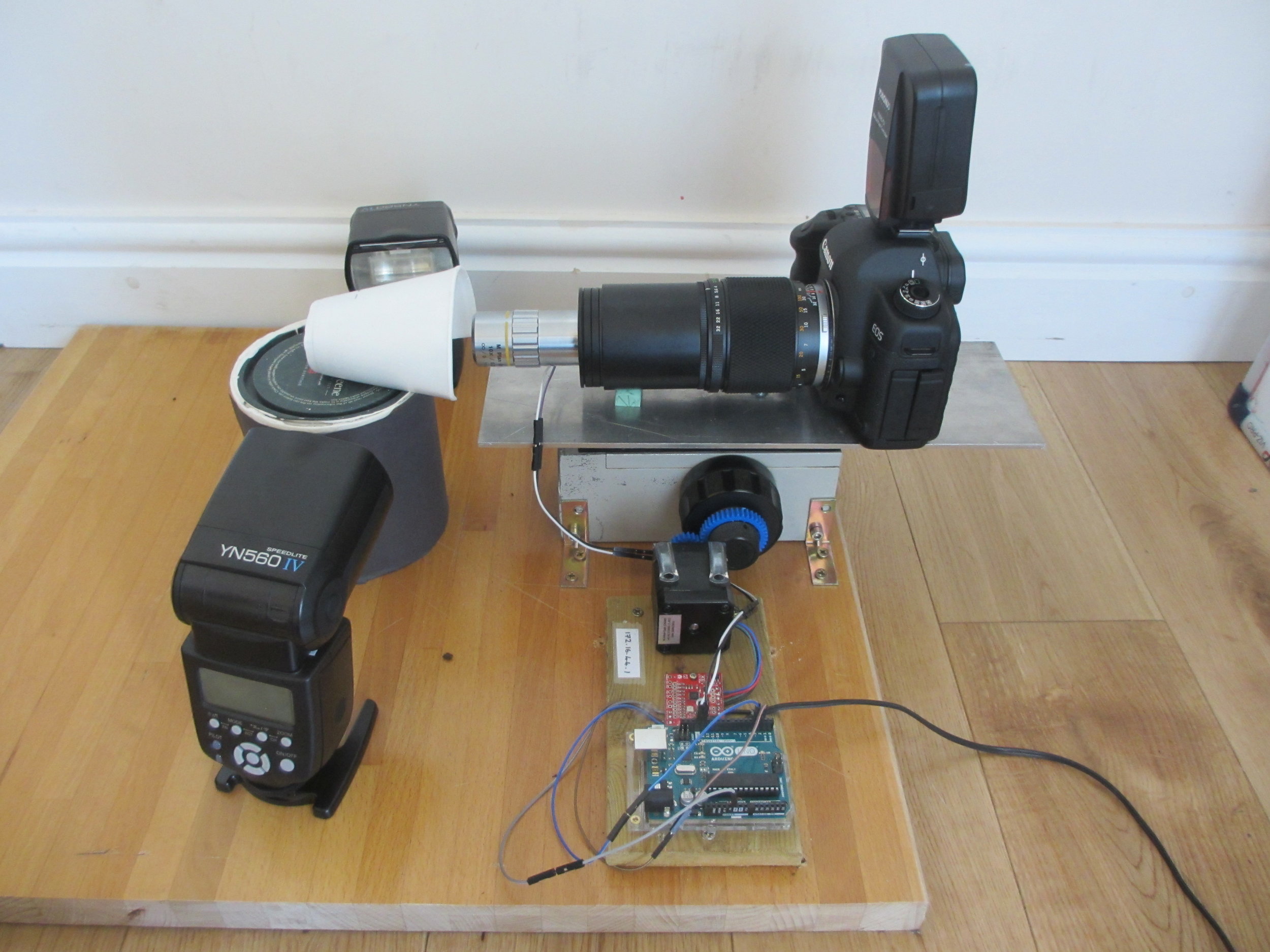
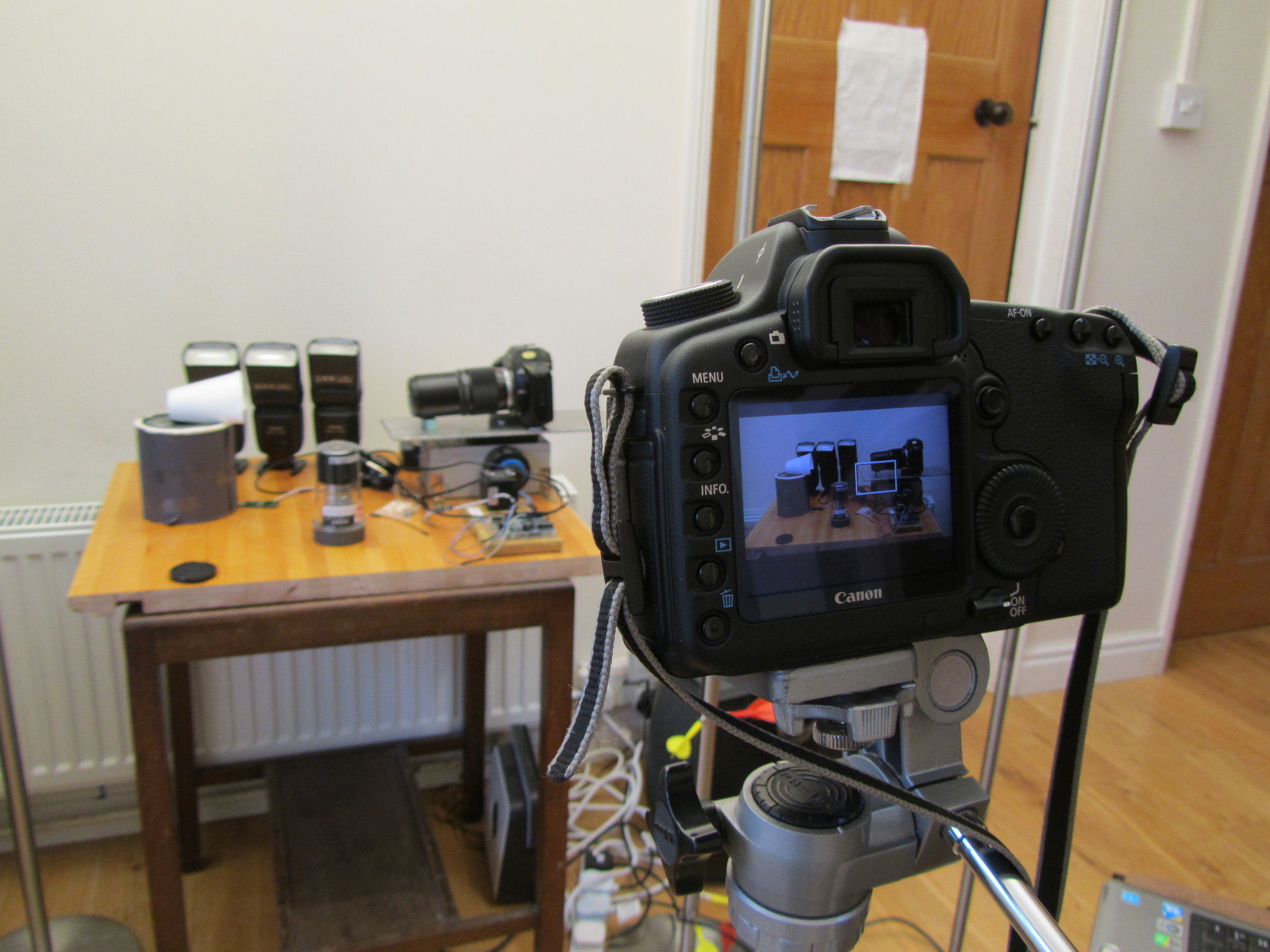
One of my funniest solved problems is that my DSLR is the only camera that I have that can record video, but it also has to appear in the videos. I got around this problem by putting my 27-year-old film SLR as a body double in the videos. The photo to the right shows my DSLR filming the focus stacking setup, with decoy camera body in place. It’s great fun editing the sound of the camera shutter into the finished video.

My other challenge is making these rather technical videos engaging to watch. There is a definite risk of them coming over as a bit dry, and so I try to keep them short and make the images interesting. I think that if I can improve my editing equipment at some point, I could make my videos much more engaging.

I’m really enjoying making educational videos and would like to keep doing this work after the end of the OpenPlant grant. I’ve been in touch with the University Public Engagement Office, who have been very helpful, and I’m hoping to learn some tips from them.


You have been awarded both a Biomaker Challenge and OpenPlant Fund grant. How have these enabled the development of the project?

JD: My work absolutely could not have been done without these grants. Most of the work has been done through collaboration, volunteer labour, and home engineering. However, the grants paid for the microscope objectives. Without these amazing lenses, I could not have done the work.


How do you feel the project is progressing?

JD: I think it's going very well. I have four good videos already online, and a lot of written documentation. I have registered a new domain (www.chlorophyllosophy.uk) as a central doorway to all of the material, and I still have lots of ideas for other videos to make.

Two out of three of my lenses have arrived and I am looking forward to taking some great photos. My Utricularia gibba (bladderwort) plants are growing well in their casserole dish. Utricularia gibba is a small, carnivorous aquatic plant that develops traps to capture its prey. They are being studied by my collaborator Christopher Whitewoods at the John Innes Centre and I have already taken my first few photos of them, as the new traps develop. The traps have a beautiful structure, and as an aquatic plant, will be a great challenge to photograph.

I hope soon also to visit the Sainsbury Laboratory in Cambridge to photograph the trichome mutant phenotypes in Arabidopsis thaliana, belonging to my collaborator Aleksandr Gavrin. I really look forward to the challenge of photographing trichomes, that will have other trichomes behind to confuse my software.

I have also just sewn a new batch of fern spores and those plants will be a real treat to photograph when the time comes.


What are the future opportunities to take this project forward?

JD: One of the biggest pitfalls for photographers is that they become so fascinated by the stream of newer and better camera equipment, that they forget to actually take any photos. I think that in the next couple of years, it's very important that I actually take the time to take some photographs. With this new technology that I have built, and with the opportunity of my volunteer labour, these will add hugely to the body of research knowledge.


Jennifer's project is also documented on Github: https://github.com/BioMakers/Gametophyte-Fern-photography-2018/blob/master/README.md

My OpenPlant Experience: Outreach, Engagement and 3D printing

Guest blog post by Roger Castells-Graells about his OpenPlant Fund project “Accessible 3D Models of Molecules”. Roger recently won a UEA Engagement Award in recognition of the work he has done both with OpenPlant and beyond.


PhD student Roger Castells-Graells in the lab

PhD student Roger Castells-Graells in the lab

My name is Roger and I am a PhD student in Prof. George Lomonossoff’s lab at the John Innes Centre in Norwich. My research project is about the production of virus-like particles to understand viral dynamics for future applications and to generate new bionanotechnological tools. I have a passion for science communication and public engagement and I have had numerous opportunities to communicate my science in Norwich, the UK and abroad since the start of my PhD.

My OpenPlant experience started in September 2016, when I attended a great Co-Lab workshop organized by the Open Science School and funded by an OpenPlant Fund. With this opportunity I had the chance to interact with scientists from different fields and also with designers and artists. It was an enriching experience and we developed a project called VRICKS (Virus Bricks) that aimed to generate tools to explain viruses in educational ways, like for example with paper models.

Following up from this workshop, in October 2016, I organized an activity for the Norwich Science Festival, together with Jenni Rant (The SAW Trust) and Colette Matthewman (OpenPlant), where we recreated the assembly of proteins into a virus protein coat using materials like paper and plastic, which represented the subunits of the virus. The public contributed to the assembly of a virus model, they learnt about related research from the Lomonossoff lab and they took home a build-at-home model. Over one hundred people participated in the activity during the weekend, making it a roaring success.

Presenting the virus activity and engaging with people at the Norwich Science Festival

Presenting the virus activity and engaging with people at the Norwich Science Festival

Following up with the interest to build tools to explain biological processes, such as virus assembly, I decided to apply for and OpenPlant Fund with the project “Accessible 3D Models of Molecules”. The project team is a multidisciplinary team (molecular biology, bioinformatics and engineering) of students from JIC and University of Cambridge and with this fund we are developing models of viruses and proteins using 3D printing technologies.

3D printed virus models for the OpenPlant Fund project
 
Normal
0


false
false
false

EN-GB
X-NONE
X-NONE


 <w:LatentStyles DefLockedState="false" DefUnhideWhenUsed="false"
DefSemiHidden="false" D…

3D printed virus models for the OpenPlant Fund project

Recently I presented some of the virus models in a high school with students aged 12 to 16 years old. The students enjoyed being able to handle and compare representations of real virus structures and were amazed that some of these structures were only discovered this year. When the school teacher was asked about how the use of educational 3D models in the classroom could benefit the learning process he answered that first of all it creates excitement and focuses the attention of the students. It is something completely new! It contributes to the understanding of three-dimensional models and gives the students a better sense of the reality of the object. Furthermore, it allows the students to calculate scale as it is possible to touch, measure and compare different models.

I was invited to speak at the Pint of Science Festival in Norwich in May, and gave a talk entitled “20000 Leagues under the microscope: Viruses & Nanomachines”. At the event, I passed around several models of 3D printed viruses and the public loved having the opportunity to handle them. It was a great experience and we received really positive feedback. I want to thank the organizers of Pint of Science for such a great event!

As a result of all of these activities, I was recently awarded a UEA Engagement Award 2016/17 for contribution to Public & Community Engagement, which I am very proud of.

  Norwich Pint of Science Festival tweets


Norwich Pint of Science Festival tweets

Roger tweets2b.png

With thanks to my supervisor Prof. George Lomonossoff, OpenPlant and all the people that have helped, encouraged me and opened up opportunities in this last year.

OpenPlant Scientists take part in Norwich Pint of Science Festival

In May 2017, the Pint of Science festival returned to Norwich. The festival, which is held over a few days, was a huge success, with many events being sold out days in advance. Each event offers the audience the chance to meet scientists at their local pub and discuss their latest research in an informal and welcoming atmosphere, whilst sipping on their favourite pint.

Two sell out events where those of OpenPlant Project Leader Professor George Lomonossoff and his PhD student Roger Castells-Graells, and a second event with OpenPlant’s Norwich-based Director, Professor Anne Osbourn.

George’s talk was entitled ‘Just Eat Your Greens – A New Way of Vaccinating?’ and took place at the York Tavern. It covered the use of a highly efficient transient expression system developed in his laboratory. This Hypertrans® system allows for the relatively quick and cheap production of large quantities of virus-like particles in plants, which have been proven to be effective as experimental vaccines.

3D printed viruses.png

Roger presented ‘20,000 Leagues Under the Microscope: Viruses & Nanomachines’ taking the audience on a journey into the nano world of viruses. During the entertaining talks, the audience took part in various activities such as making a virus molecule out of pipe cleaners and creating virus inspired sketches on beer mats.


The following evening, Anne took to the stage at the St Andrews Brewhouse to present her ‘Finding Drugs in The Garden’ talk. Anne’s inspiring talk invited people into the plant kingdom to hear about its very own chemistry toolkit. She presented her teams current work harnessing the DNA that encodes the pathways to these chemicals and using them to produce designer molecules for medicinal, agricultural and industrial applications.

water-2986837_960_720.jpg

For the scientists taking part in the festival, it has proven to be a great platform on which to reach the public to talk about their research and build an understanding of their work within the local city of Norwich. After such well received talks and events, we very much look forward to the return of the Pint of Science Festival in 2018.

OpenPlant Fellow wins 2016 Wellcome Trust Image Award

fernan-maize Fernán Federici, OpenPlant Fellow at the University of Cambridge and Director of the Synthetic Biology Lab at Pontificia Universidad Católica in Chile has been awarded a Wellcome Trust Image Award for his micrograph of maize leaves, shot in the Department of Plant Sciences in collaboration with Professor Jim Haseloff.

Fernán has enjoyed considerable success with his artistic images of bacteria and plants using microscopy, winning Wellcome Trust awards in 2011 and 2012. His image has appeared in multiple media outlets, including Nature Newsthe BBC and The Guardian.

More on the image via the Wellcome Trust >>

Looking inside a cluster of leaves from a young maize (corn) plant reveals lots of details and organised structure. Each curled leaf is made up of lots of small cells (small green square and rectangle shapes), and inside each cell is a nucleus (orange circle), the part of the cell which stores genetic information. Maize is one of the most widely grown cereal crops in the world. It is used as a staple food, in livestock feed, and as a raw material – such as for processing into high-fructose corn syrup. Genetically modified maize crops are being grown to be resistant to pests and herbicides.

Although seeming boring when viewed with the naked eye, maize leaves have such a delicate and intricate structure under the microscope, captured so wonderfully by this picture. The level of detail as demonstrated by the image reminds us how complex even relatively simple organisms are when seen on this scale.

James Cutmore, Picture Editor of BBC Focus

The image has been made freely available to use under a Creative Commons CC-BY-NC-ND 4.0 license.

OpenPlant lab mentioned in 'Microalgae as a microcosm of plant biotech'

Logo-monserrat chlamydomonas photo

Source: Microalgae as a microcosm of plant biotech

Orlando de Lange has a blog post up on 'Microalgae as a microcosm of plant biotech' mentioning the work of Alison Smith's Lab at the University of Cambridge, plus several others using Chlamydomonas for classical biotechnology and synthetic biology. Worth a quick read!

And of course I won’t leave out synthetic biology. Several labs seem to be exploring the potential for synthetic biology with microalgae.Alison Smith’s lab in Cambridge has long studied mircroalgal metabolism, with an eye to biofuel production and has more recently begun churning out tools for engineering Chlamydomonas.

Plant Synthetic Biology at IHÉS

poster_cellular_molecular Institut des Hautes Études Scientifiques (IHÉS) held a meeting on Cellular and Molecular Biotechnology, including Synthetic Biology in Paris last week. OpenPlant was well represented by Professors Jim Haseloff and Anne Osbourn. You can find videos of their talks below and all recordings on the IHÉS youtube channel.

https://www.youtube.com/watch?v=n5Yjj3hMtZo

https://www.youtube.com/watch?v=xSKM1YcM4rs

https://www.youtube.com/watch?v=9FN6E1xTAic