OpenPlant Blog — OpenPlant

Sami Stebbings

Publication: Discovery of putative Golgi S-Adenosyl methionine transporters reveals the importance of plant cell wall polysaccharide methylation

OpenPlant PI Professor Paul Dupree recently published work in collaboration with colleagues from the University of Cambridge, The Andrés Bello National University, University of Warwick and Massachusetts Institute of Technology describing the identification of putative transporters that import SAM into the Golgi lumen in plants, providing new insights into the paramount importance of polysaccharide methylation for plant cell wall structure and function.

Henry Temple, Pyae Phyo, Weibing Yang, Jan J. Lyczakowski, Alberto Echevarría-Poza, Igor Yakunin, Juan Pablo Parra-Rojas, Oliver M. Terrett, Susana Saez-Aguayo, Ray Dupree, Ariel Orellana, Mei Hong, Paul Dupree

https://www.biorxiv.org/content/10.1101/2021.07.06.451061v1

Abstract

Polysaccharide methylation, especially that of pectin, is a common and important feature of land plant cell walls. Polysaccharide methylation takes place in the Golgi apparatus and therefore relies on the import of S-adenosyl methionine (SAM) from the cytosol into the Golgi. However, to date, no Golgi SAM transporter has been identified in plants. In this work, we studied major facilitator superfamily members in Arabidopsis that we identified as putative Golgi SAM transporters (GoSAMTs). Knock-out of the two most highly expressed GoSAMTs led to a strong reduction in Golgi-synthesised polysaccharide methylation. Furthermore, solid-state NMR experiments revealed that reduced methylation changed cell wall polysaccharide conformations, interactions and mobilities. Notably, the NMR revealed the existence of pectin ‘egg-box’ structures in intact cell walls, and showed that their formation is enhanced by reduced methyl-esterification. These changes in wall architecture were linked to substantial growth and developmental phenotypes. In particular, anisotropic growth was strongly impaired in the double mutant. The identification of putative transporters that import SAM into the Golgi lumen in plants provides new insights into the paramount importance of polysaccharide methylation for plant cell wall structure and function.

Vacancy: Postdoctoral Research Associate

Postdoctoral Research Associate, Developing Forecasts of Environmental Risk to Carbon Stocks in Nature-based Systems, Forest Ecology Group (Coomes Lab), Cambridge Centre for Carbon Credits (4C), University of Cambridge, Cambridge UK.

The Position

You will help to develop a standardised approach to forecast the expected permanence in carbon stocks of nature-based projects, as influenced by human activities, climate change, and other disturbance events. You will stochastically extend an existing deterministic difference-in-difference analysis so that it can be used to estimate the expected permanence of nature-based carbon stores and credits derived from them.

The modelling will involve combining a variety of different risks, from climate change to the socio-economic drivers of deforestation, and how these are modified through project interventions. Your work will necessitate foundational work on risk-modelling and its application to our automated system for generating and valuing carbon credits.

Salary: £33,309 - £40,927

Application deadline: 5 April 2022

More information on this position can be found here.

Vacancy: Postdoctoral Research Associate

Assessor of Carbon Sequestration by Nature-based Climate Solutions, Forest Ecology Group (Coomes Lab), Cambridge Centre for Carbon Credits (4C), University of Cambridge, Cambridge UK.

The Position

The successful candidate will join the Cambridge Centre for Carbon Credits (4C) which aims to verify the claims and consequences of carbon credit generating activities, to ensure these have the most beneficial impact as we transition to a net-zero future.

You will assess and track changes in carbon stocks working with other research groups and private sector partners. You will bring together appropriate, trusted primary observations from ground-based surveys with remote sensing datasets to measure and track carbon stocks through time. The contexts would include key ecosystems, such as forests and peatlands. The data products should scale to the global level and will be used as an input to our automated system for generating carbon credits.

Salary: £33,309 - £ 40,927

Application deadline: 5 April 2022

More information on this position can be found here.

Churchill Fellowship

The Churchill Trust is proud to announce that a new Churchill Fellowship will be offered from 2022 to honour the memory and legacy of the late Saskia Beer, made possible through the generous sponsorship of Colin and Maggie Beer.

Churchill Fellowships are an internationally recognised award, providing access to expertise from around the world. Applicants are empowered to design their own projects, enabling them to explore international best practice and innovation that can be applied in Australia. There is a high level of visibility and credibility associated with becoming a Churchill Fellow, as well as a responsibility to share the knowledge and skills gained overseas with the Australian community. No prescribed qualifications are required to apply for a Churchill Fellowship and the subject of proposed projects is limitless, provided there is evidence of a benefit to Australia.

Application deadline: 28 April 2022

More information about this fellowship can be found here.

EFSA Traineeships Call 2022

The European Food Safety Authority (EFSA), based in Parma, Italy is opening positions for trainees.

The position

Under the supervision of your traineeship adviser within the unit of assignment, you will have the opportunity to:

  • Get a first-hand experience into the work of a scientific EU Agency, developing new competencies and skills in line with your personal interests and professional aspirations;

  • Contribute to the overall objectives of the unit by supporting in drafting documents and reports, handling scientific and/or administrative work, and ensuring timely communication of outcomes;

  • Contribute to specific projects and activities with new ideas and innovative solutions;

  • Contribute to the planning and monitoring of the unit’s activities;

  • Participate in several trainings which will help you grow professionally.

Application deadline: 25 April 2022

More information about the position can be found here.

Vacancy: Bioinformatician

Applications are invited for a Bioinformatician to join the Laboratory of Dr Falk Hildebrand at the Earlham Institute.

The role:

This post requires an experienced bioinformatician with an interest to study reconstructed microbial genomes in metagenomes (bacterial, archaeal, fungal). The post-holder will be involved in conceptualizing & implementing bioinformatic pipelines to analyse large time-series metagenomes (human gut, soil, …), based on data from 2nd and 3rd gen sequencing platforms, such as illumina, PacBio, ONT, Hi-C & single cell sequencing data. The analysis will be published in research papers that the candidate should be leading.

Salary range: £32,578 - £39,938

Application closing date: 14 April 2022

More information about this position can be found here.

OpenPlant and the SAW Trust launch a new synthetic biology resource for schools

Synthetic Biology is seen as an emerging and important multidisciplinary field, that is currently under-represented in the national curriculum and teaching resources. Collaborating with the Science, Art and Writing (SAW) Trust, researchers from across OpenPlant have produced SynBio 4 Schools; a free to use, comprehensive package of educational resources that focuses on teaching the principles of plant synthetic biology for GCSE and A Level students.

SynBio4 Schools

Inspiring the next generation of biological engineers.

The new resource was officially launched last month with the hosting of a live, online launch event. Teachers were invited to join the event and ask any question they might have on delivering the resources and were given the opportunity to hear more about the science behind the resource activities from some of the scientists who created them; Professor Julea Butt (University of East Anglia), Dr Nicola Patron (Earlham Institute), Dr Roger Castells Graells (University of California, LA) and Dr Shannon Woodhouse (John Innes Centre).  Dr Paolo Bombelli, who also created a resource recorded a step–by-step video on how to build a plant microbial fuel cell to compliment the set.

SynBio 4 Schools is comprised of a downloadable booklet, which covers an introduction to synthetic biology and its applications, step-by-step guides to 4 practical activities for schools to conduct and a supporting teacher notes booklet which highlights the relevance of each activity to the British school science curriculum.  There are also two videos; the previously mentioned ‘Build Your Own Plant Microbial Fuel Cell’ with Paolo Bombelli, and four introductory videos to each of the SynBio 4 Schools Activities.

Topics covered include; Building DNA Circuits, Generating Electricity From Plants, What Can Viruses Do For You? and Natural Product Synthesis. It is hoped that these activities will help schools engage more with synthetic biology practises, whilst inspiring the next generation of biological engineers.

All resources are available via the OpenPlant website and are free to use.

Vacancy: Postdoc Position - Plant Molecular Biology

Postdoc Postion, Elina Lab, Crop Science Centre, Cambridge

A post-doctoral position in Dr Natasha Yelina’s lab working on meiosis in model and crop plants at the Crop Science Centre, University of Cambridge.

Closing date: January 23rd.

For more information visit the University of Cambridge website.

https://www.jobs.cam.ac.uk/job/32757/

Independent Research Fellowships

The John Innes Centre are welcoming applications from who either hold, or wish to apply for Independent Research Fellowships (such as a UKRI Future Leaders Fellowship, or a Royal Society University Research Fellowship).

Shortlisted candidates will be invited to give a seminar at the Fellows Conference, which will be held on 7 February 2022.

To apply email a two-page summary of your research plan, a copy of your CV and arrange for three letters of recommendation to be emailed to fellows@jic.ac.uk

Closing date December 3rd 2021

For more information - https://www.jic.ac.uk/vacancies/independent-research-fellowships/

sciTAG: a label design and printing app created by scientists for scientists

Proper labelling practices in the lab is one of the first things students are taught at the practical courses. As a matter of fact, labelling is an essential part of data management for scientists while working in the laboratory. And if you ever worked in a lab, you must know how much time you usually spent to label every single tube, falcon, vial, bottle etc.

The Problem

Appropriate labelling is an essential research practice. It is fundamental to day-to-day operation and long-term sample storage in laboratories. Creating labels with handwriting is convenient, adaptable, and often the standard method scientists use. However, variabilities in label format and illegible messy handwriting can both lead to flawed and/or erroneous communication, resulting in, for example, loss of important biological samples. Moreover, writing labels by hand for large scale experiments or a high number of samples is time-consuming and creates ergonomic stress. The problem presented called for a labelling method that is automated, user-friendly, and affordable.

The Project

We proposed to develop an IOS app that allows biological and medical laboratories to operate in a consistent and efficient manner, by making label design and printing more accessible.

We envisaged an app which is user-friendly, automated, and compatible with affordable label printers. To accomplish our goal, we followed a stepwise approach composed of three parts: user interface design, technical structure of the app and testing the app.

Efficient label creation frees scientists from the laborious task of writing labels by hand. Consistently and systematically designed labels ensure samples stored for long term are readable and used accurately. The app will be free, meaning that convenient label printing will be accessible to the wider scientific community.

What Has Been Achieved So Far?

Our team of five is composed of a molecular biologist, a microbiologist, a geneticist, a bioinformatician and a software developer. We started by self-teaching ourselves how to design and develop an app since most of us never worked on app development before.

Interface design decisions were made not only to be attractive to potential users, but also to be functional and simplistic as possible. One aspect of the design is visual elements. We decided on aesthetic choices such as a colour scheme throughout the app. We considered colour blindness since 4.5% of the global population experience colour-blindness. Next, we decided on the name of the app: sciTAG. We did market research to make sure this name is not a trademark. Finally, we designed an app logo depicting a double-stranded DNA alongside the name of the app.

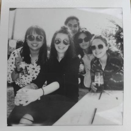
From left: Junyan Liu (Postdoctoral Researcher, Sanger Institute), Marta Matuszewska (PhD student, Department of Veterinary Medicine, University of Cambridge) Begum Akman (Research Associate, Department of Pharmacology, University of Cambridge), Chuqiao Gong (Software developer, EMBL-EBI), Ellis Kelly (PhD student, Department of Genetics, University of Cambridge)

Then, we identified the features that are crucial for our target audience. We divided the label design into two blocks by keeping it as simple as possible with choices included to create a good label.

From left to right: home screen, label design screen, prototype label in PDF format.

Next step was to layer the technical structure: first user input information used to create a printable object such as PDF. Second, to convert this object to a storable object within the app. Finally, the app had to be compatible with commercially available label printers.

What’s Next For The sciTAG Team?

With the help of the Biomarker Challenge funding, we sourced several label printers available in the market to test sciTAG app. Among these printers only one of them is specific for lab settings (very expensive!) and others are highly affordable printers that we sourced from different companies. We distributed these printers within the team, and we are planning to complete the sciTAG prototype, start testing and reporting our experiences.

After the successful tests from the team, we are planning to recruit five laboratories within University of Cambridge to further test and optimize the sciTAG app. Eventually we are aiming to make sciTAG freely available to the science community on the App Store. Further down the line we also would like to consider making our app available for android users.

Synthetic Biology UK 2021

Synthetic Biology UK 2021 will be hosted by Synthetic Biology Research Centre Nottingham, and will focus on a number of burgeoning new areas of activity, including the engineering of microbial communities, microbial-plant interactions and applications for the sustainable manufacture of materials, the circular economy and climate mitigation strategies.

Date: November 22nd

For more information and to register visit:

https://www.eventsforce.net/biochemsoc/frontend/reg/thome.csp?pageID=53019&eventID=108&CSPCHD=0000010000005HOPapInI12EMFR969aLR0xa4Zi1NHfz6Lg5Pw

Vacancy: Laboratory Manager at Leaf Expression Systems

Leaf-Expression-Systems.jpg

The Company

Leaf Expression Systems is a contract development business based in Norwich, Norfolk which specialises in the plant-based expression and production of proteins, metabolites and complex natural products for research and biomedical applications using a proprietary, transient expression technology, Hypertrans®.

The role

Position type: permanent

Hours: Full time

The Laboratory Manager will oversee the efficient day-to-day running of Leaf Systems laboratories, growing areas and general facilities, providing a range of services in support of the company’s development and manufacturing activities. The hands-on role will ensure that the technician team are maintaining all general laboratory supplies, sufficient plants are available for production demand and perform waste management duties, which includes using autoclaves. The Laboratory Manager will also ensure that all environmental health and safety requirements are met by the team and quality management documentation is followed.

For more information go to www.leafexpressionsystems.com/careers/

All applicants must apply with a covering letter and CV to info@leafexpressionsystems.com

Stress priming for production of biodiesel precursors

The aim & rationale

Unicellular algae form a powerful biotechnological warehouse for production of chemical compounds and a valuable platform for synthetic biology. However, algae commercial potential can be fully revealed only after deepening the understanding of their fundamental processes. Triacylglycerols (TAGs) are lipids that can be used as the precursors in biodiesel production. Model green alga, Chlamydomonas reinhardtii, accumulates TAGs in stress conditions, like nitrogen deprivation. TAGs accumulation is reversible and chromatin-based, features that make this process a potential subject for stress priming - a phenomenon where repeated stress exposure leads to amplification of the response comparing to the 1st/single stress exposure (conceptually similar to vaccination). Stress priming was not explored in algae, even though it offers a tool to manipulate algal biofuel production!

We proposed to establish and optimize protocols for algal growth and TAGs’ accumulation in stress priming setup using a histochemical, chromatographic and microscopic approach. The aim of the project was to decipher whether stress priming exists in simple eukaryotes and whether it can be used for biotechnological/synthetic biology purpose.

The project

We undertook the following steps: 1) Set up stress priming growth conditions, including the selection of proper harvesting timepoints and preservation of harvested material; 2) Implement and optimize simple TAGs staining protocol using Nile Red dye; 3) Acquire images using confocal microscope; 4) Quantify dye intensity, normalize to cell size and extrapolate to TAGs accumulation strength (Nile Red fluorescence is tightly correlated with TAGs content); 5) Obtain chromatographic profile of accumulated TAGs using GC-MS.

Figure 1: TAG-stained C. reinhardtii cells with Nile Red (green/yellow). Red corresponds to the autofluorescence.

Figure 1: TAG-stained C. reinhardtii cells with Nile Red (green/yellow). Red corresponds to the autofluorescence.

After some optimization, we found out that some fiddling around with recovery time, staining protocol and extraction are needed (see the link to our Hackster page below), but overall, the plan worked. We obtained some beautifully stained TAGs in alga cells and sought to properly quantify fluorescence intensity with normalizations to account for the background noise and batch variation. Finally, we were able to properly compare stress-primed samples and single-stress controls. Long story short, we saw that… stress priming indeed occurred in the conditions used for TAGs’ accumulation in model green alga! However, to our surprise the dynamics were opposite to expected – stress priming caused TAGs’ downregulation, instead of upregulation!

We wanted to study the phenomenon further and understand lipid type composition in our samples. To this end, we employed gas chromatography. Despite quantitative difference in TAGs accumulation, stress-primed cells did not show qualitative difference in lipid type profile. However, we did observe that stress samples in general exhibited lower lipid complexity than non-stressed controls.

Future outlook

Our results show that stress priming exists in simple alga. Since stress treatment is frequently employed for biosynthesis of valuable bioproducts like biofuel, antioxidants or nutraceuticals, this phenomenon can be used for biotech & synthetic approaches.

As for direct outcomes, our Hackster page contains protocols and descriptions for procedure optimization, that can be used by DIY biotechnologists. We are also drafting a small paper for the broader community. Alga growth and staining procedures can be performed in a cost-effective manner and expensive equipment (we admit we used it) can be replaced by cheaper alternatives developed in other Biomaker projects (DIY microscopes and bioreactors). The Biomaker challenge opened us to new ideas and is worth a recommendation to all the curious people!

Acknowledgements

We would like to thank the organizers for making this experience happen and Andy Truman (John Innes Centre, Norwich) for being our kind host of the grant.

Please check out our hackster page to learn more about the project.

Pawel Mikulski and Javier Santos Aberturas, John Innes Centre.

The Global Garden project: Imagining plant science

The Global Garden project: Imagining plant science

The Global Garden project: Imagining plant science.

Nicholas M. Lee, Hannah E. Hodgson, Chris Hann, Mike O’Driscoll, Samantha Stebbings, Collette Matthewman, Miriam Kent, Jenni Rant and Anne Osbourn

Plants, People, Planet (2020) 2: 602-613

https://doi.org/10.1002/ppp3.10133

Publication: Comparative analysis of early divergent land plants and construction of DNA tools for hyper-expression in Marchantia chloroplasts

Publication: Comparative analysis of early divergent land plants and construction of DNA tools for hyper-expression in Marchantia chloroplasts

Comparative analysis of early divergent land plants and construction of DNA tools for hyper-expression in Marchantia chloroplasts.

Eftychios Frangedakis, Fernando Guzman-Chavez, Marius Rebmann, Kasey Markel, Ying Yu, Artemis Perraki, Sze Wai Tse, Yang Liu, Jenna Rever, Susanna Sauret-Gueto, Bernard Goffinet, Harald Schneider and Jim Haseloff.

BioRxiv (2020) 2020.11.27.401802

https://doi.org/10.1101/2020.11.27.401802

Publication: A GRF-GIF chimeric protein improves the regeneration efficiency of transgenic plants

Publication: A GRF-GIF chimeric protein improves the regeneration efficiency of transgenic plants

A GRF-GIF chimeric protein improves the regeneration efficiency of transgenic plants.

Juan M Debernardi, David M Tricoli, Maria F Ercoli, Sadiye Hayta, Pamela Ronald, Javier F Palatnik, Jorge Dubcovsky.

Nature Biotechnology (2020) 38: 1274–1279

https://doi.org/10.1038/s41587-020-0703-0

Publication: Formation and diversification of a paradigm biosynthetic gene cluster in plants

Publication: Formation and diversification of a paradigm biosynthetic gene cluster in plants

Formation and diversification of a paradigm biosynthetic gene cluster in plants.

Zhenhua Liu, Jitender Cheema, Marielle Vigouroux, Lionel Hill, James Reed, Pirita Paajanen, Levi Yant and Anne Osbourn.

Nature Communications (2020) 11: 5354.

https://doi.org/10.1038/s41467-020-19153-6

CtoD: From Cells to Droplets

The aim and rationale

Real biological systems are complicated by the massive number of components and highly complex cellular network. Establishing a cell-like environment in vitro would allow us to disentangle basic mechanisms underlying cellular networks under well-controlled settings. We are a small but interdisciplinary team between the Department of Chemistry and the Department of Plant Sciences, aiming to apply microfluidic droplet system to studying physiological processes in plants, transforming the objects of research from plant tissues into artificial cells.

CtoD Betalain enzyme.png

Microdroplets generation and in-droplet protein expression for mimicking physiological pathways.

The project and more

In this project, we aimed to develop an artificial cellular system using microfluidic droplets that allows the in vitro expression of proteins from plants in a compartmentalised cell-like environment. The biosynthetic pathway of a class of plant pigments, betalains, was used as a simple model to test the methodology, and the TNT® SP6 High-Yield Wheat Germ Protein Expression System from Promega was selected as a reliable in vitro protein expression system. The cell-free protein expression components together with betalain biosynthetic genes were enclosed in microdroplets and intermediate and final products measured.

Betalain biosynthetic pathway from L-Tyrosine to two groups of betalains. DOPA, 3,4 dihydroxyphenylalanine. DODA, 4,5 DOPA dioxygenase. cDOPA5GT, cyclo-DOPA 5-O glucosyltransferase.

Betalain biosynthetic pathway from L-Tyrosine to two groups of betalains. DOPA, 3,4 dihydroxyphenylalanine. DODA, 4,5 DOPA dioxygenase. cDOPA5GT, cyclo-DOPA 5-O glucosyltransferase.

Up to this point, we have tested the expression of various proteins in microdroplets. Betalain biosynthetic enzymes were produced and confirmed by fluorescent protein fusions in microdroplets. However, simply co-expressing enzymes from the betalain biosynthetic pathway in the cell-free protein expression solution was not sufficient to drive detectable production of pigment molecules. Following these, optimisation of buffer composition was made and various substrates fed into the system, which led to realisation of parts of the pathway in the in vitro environment.

In addition to the project itself, the various events and training organised by OpenPlant/Biomaker were even more remarkable. The annual OpenPlant Forum gathered an incredibly diverse range of subjects and the low-cost DIY instruments for automating laboratory experiments were mind-blowing.

Future perspective

The OpenPlant/Biomaker project has built solid foundation and connection for us to move forward in the future. Communication between cells plays a central role in many metabolic pathways of multicellular organisms, yet it remains difficult to reproduce in vitro. To this end, an artificial multicellular system with droplet-to-droplet communication will be explored to mimic metabolic pathways which rely on the communication between cells. This platform would open more opportunities for synthetic biology and allow hypotheses and models based on cell-to-cell communications to be tested in vitro.

Last but not least, we thank the OpenPlant/Biomaker teams for the wonderful events and generous help that they have provided!

Written by Zhengao Di, University of Cambridge


Developing an open and affordable 3D bioprinter

Background

Friends had Central Perk cafe, the gang from How I Met Your Mother had MacLaren’s Pub and we have Charlie’s Pizza joint. What started as a trivial discussion about printing human organs over a stone-baked Margherita quickly evolved into us applying for a bid to participate in the BioMaker Challenge, with the objective of Developing an open & affordable 3D bioprinter

The beginnings of 3D printing date back to the 1980’s, when it was commonly known as its more mouthful synonym stereolithography (SLA), a technology pioneered by Charles W. Hull. Revolutionized with SLA, one could translate a 3D design from a data file into a physical object, in a relatively short time. The household notion of 3D printing is testament to the success of SLA, which originated from the company 3D Systems Corporation. 

Fast forward 40 years, my neighbours are at home 3D printing little elves for their garden, my cousin is buying a 2-story building printed by an enormous SLA machine, my doctor is offering his patients custom 3D printed ear implants, heart valves or bones - SLA has already infiltrated many aspects of our lives, economy and medical care. Much of this progress came about because of the RepRap project, whose aim was to create an open-source 3D printer capable of printing most of the parts needed to replicate itself, making it cheap enough for hobbyists. Today’s consumer 3D printer companies, and the widespread use of 3D printing, grew out of the RepRap hobbyist community. 

The field of regenerative medicine could hugely benefit from 3D printing techniques to offer personalized medicine to its patients, by for example printing organs made of one’s own cells. However, these applications are challenged by the complex architecture of human organs, the difficulties of supporting living cells, introducing foreign materials in a human body and working at a microscale. 

Addressing these issues, companies and academic researchers have built 3D printing platforms, specialized for biological materials, hence 3D bioprinting. Not surprisingly, these come at a prohibitive cost, up to £200K for the printer only.

BioMaker Challenge

During our weekly pizza gatherings we talked about how it would be great to have an open-source bioprinting project, along the same lines as the original RepRap project. It could make the technology more accessible and bring printing human organs for transplantation a step closer. None of our backgrounds were in bioprinting, but our combined skillsets did range from engineering and programming to cell biology, with diverse backgrounds in start-ups, industry and academia. The Biomaker challenge would be a great way to work together on a project where we could contribute something to the 3D printing community, and learn a lot along the way. Plus it would be fun! 

3D bioprinter.

Bertie the 3D bioprinter.

So as part of the BioMaker Challenge, we set out to address the lack of reasonably priced open-source 3D bioprinters in the market by Developing an open & affordable 3D bioprinter. With this project still in its infancy, working with human cells was out of the scope due to ethical and practical reasons. Nevertheless, we attempted to design our 3D printer to potentially print corals, an application with a shorter timeline, more amenable to working in the absence of a biological laboratory and with direct benefits to a problem close to our hearts, the depreciation of the ocean’s corals. Our project branched into two components outlined below: 

1. Converting an existing open-source 3D printer into a 3D bioprinter capable of extruding biomaterial.

Our inspiration to address this first problem is drawn from a paper by an American group (Push et., 2018), in which they describe the design of a syringe pump large volume extruder (LVE) of low cost, compatible with printing biomaterial. This involves the purchase of standard materials (e.g: 60mL syringe, bolt nuts) and the 3D printing of other assembly components with a PLA extruder from a standard 3D printer. The newly assembled LVE would then replace the printer’s standard PLA extruder and allow the printing of biomaterials. 

With this in mind, many hours of research and discussions went into selecting the right 3D printer as a foundation for our bioprinter - we were certain about two things: it had to be open-sourced and stay within a reasonable budget. We finally settled on the RepRap Ender 5 3D-printer. In addition to the syringe extruder, we were interested in trying to integrate a peristaltic pump in our system because it would allow us to extrude multiple bio-inks at once. 

Adding a valving architecture between the reservoirs and the pump enables the switching of the reservoir that bioinks are aspirated from on the go, thereby enabling the interlinking of multiple inks of different cell types in 3D space. 

After a few months of putting all the hardware together and with a sprinkle of software, Bertie the Bioprinter was born!

2. Developing a bioink suitable for sustaining living cells.

Regardless of the organism in question, a bioink should: 

a) Contain the right nutritional environment for specialized cells.

b) Maintain a single-cell suspension (prevent cells from clumping together and/or from dying from being separated from their friends).

c) Provide a physical scaffold once printed to keep the shape of the 3D model.

d) Use materials that are safe to insert in the receiving environment. 

Printing human cells was out of the scope of Biomaker, so we initially turned our attention to another challenging bioprinting application, trying to 3D print coral (which not all of us knew was a living organism!). Coral numbers and health have declined rapidly as a result of environmental conditions and their slow growth cycle. If coral could be 3D bioprinted, perhaps it could be transplanted back into reefs to bring them back to life.

We dove into the scientific literature and identified two such bioink formulas, which are, in theory, capable of supporting the life cycle of corals and providing an “ocean-friendly” physical scaffold. It turned out however that coral species are high maintenance, requiring expensive aquariums and daily care. Even the most experienced coral growers find it challenging to maintain a community of the families of coral that were the right size to fit our bioprinter. We realised that we didn’t have the funds or the time to manage coral during the Biomaker Project, but we did make connections with coral researchers who we hope to work with on this project in the longer term. 

Bertha.jpg

Printed phytoplankton using Bertha the bioprinter.

We were however determined to evaluate the efficacy of our bioinks, and didn’t let coral’s needy demands deter us. One of the bioinks we identified is based on the chemical interaction of sodium alginate and calcium chloride, which create a solidified gel upon contact. At the concentration used, these chemicals didn’t affect the survival of phytoplankton, a single cell organism we chose to work with for its relative low cost, ease of maintenance and ability to survive as single cells. 

By the end of the project we were able to print a very slimy collection of phytoplankton, and affectionately named it Bertha. Bertha still sits in someone’s fridge for posterity.

Future

The BioMaker Challenge was an incredible opportunity for us to concretise an idea and start along the path to our long term goal. We also got to meet a wonderful community of makers and with their help and feedback we hope to further refine Bertie the Bioprinter. Moving forward, we plan to improve Bertie’s performance in terms of printing precision and its ability to run complex modes (e.g: multiple bioinks printing at once), as well as make him suitable for sterile work (e.g: ventilation, UV lights). We also plan to develop  application specific bioinks for Bertie to print. 

We’d like to take this opportunity to thank the BioMaker Organisers (special thanks to Alexandra, Jenny & Jim) for arranging this event, Professor Ludovic Vallier (Cambridge Stem Cell Institute, Jeffrey Cheah Biomedical Centre) for supporting our ideas and administrative needs as well as the BioMakeSpace Cambridge who welcomed us in their community.  Thanks to the BioMaker Challenge, we are now one small step closer to 3D printing organs, and in the meantime, Bertie is on standby for printing COVID-19 related materials (with the original PLA extruder) if need be. 

Julie, Sebastian, Monica, Robin, Ben & Tejas

PS, For more details of the project, check out our Hackster :-)